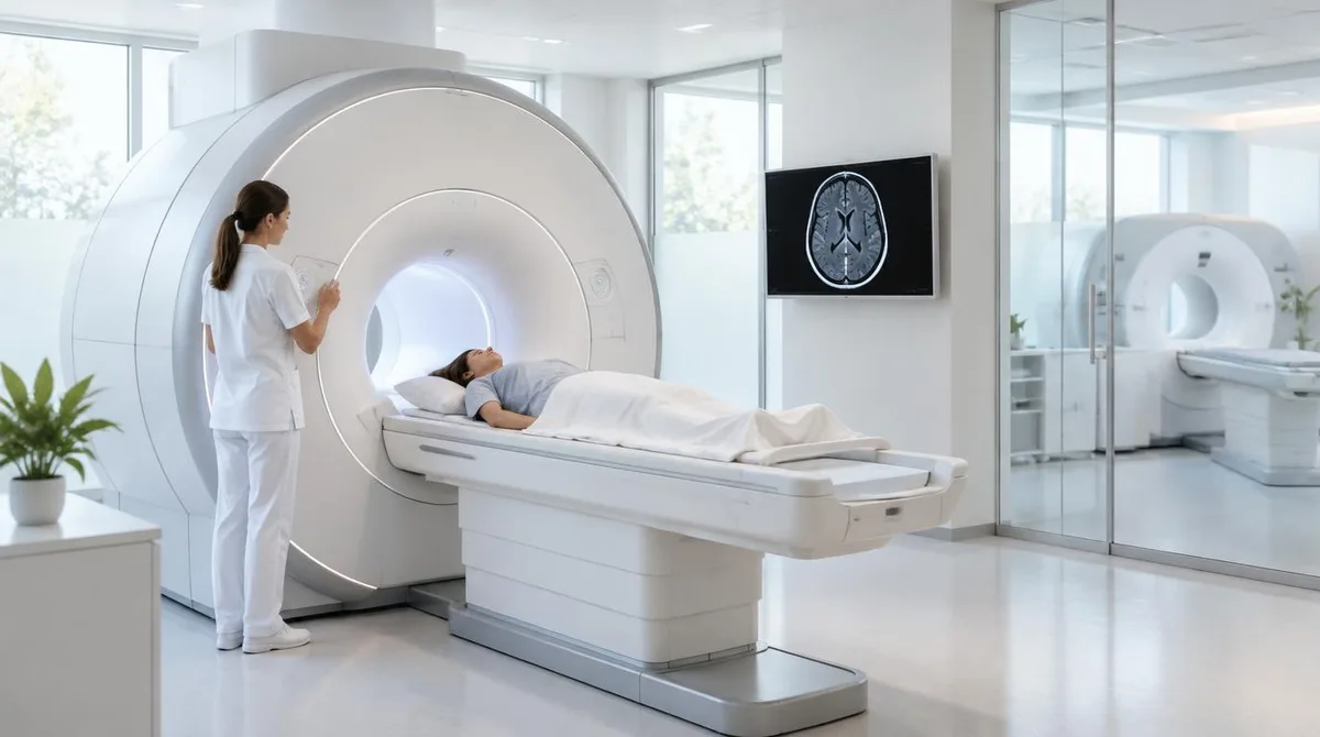
A CT scan and an MRI both show the inside of the body, but they measure different things. A CT scan uses X-rays and is fast. It gives clear pictures of bone, bleeding, and chest or abdominal emergencies. An MRI uses a strong magnetic field and radio waves, with no ionizing radiation, and gives far more detail in soft tissue such as the brain, spinal cord, joints, and organs.
Which one a doctor orders depends on the clinical question, not on which machine is "better." A neuroradiologist at MD Anderson Cancer Center put it plainly: the two scans are complementary, because they give different types of information. CT is better at spatial resolution — showing where one structure ends and another begins. MRI is better at contrast resolution — showing the difference between tissue types, which is how abnormal tissue stands out from normal tissue.
How each scan actually works
A CT scanner, sometimes called a CAT scanner, is a form of X-ray imaging. The patient lies on a table that moves through a large ring. X-ray beams hit the body from multiple angles, and a computer reconstructs them into cross-sectional images. The scan itself often takes a minute or less, and the machine is quiet. As Healthline describes it, CT scans produce detailed images of bone and less detailed images of soft tissue.
An MRI works on a different principle entirely. A powerful magnet aligns hydrogen atoms in the body's water and fat molecules. Radio pulses then knock those atoms out of alignment, and the signals they emit as they settle back are translated into an image. Because the signal depends on tissue chemistry, MRI can distinguish fluid, fat, and abnormal growths with high detail. The trade-off is time: an MRI typically runs 20 to 90 minutes, and the patient must lie very still or the affected images get repeated. For related coverage, see Memory Care Facilities: What They Are and How to Evaluate Them.
Both machines require the patient to lie on a movable table that passes through a donut-shaped opening. That is roughly where the resemblance ends.
What each test is best at
CT is the emergency tool. It is faster, more widely available, and usually less expensive. Typical uses include trauma, suspected internal bleeding, bone fractures, bowel blockages, blood clots, appendicitis, kidney stones, and lung problems. CT is also the standard first scan for stroke, and it is often used to locate tumors and to monitor whether cancer has spread. In cancer care, CT is standard for initial staging, while MRI adds detail for brain, liver, prostate, and soft-tissue tumors.
MRI is the detail tool. It is the first choice for most soft-tissue questions: brain tumors, multiple sclerosis, spinal cord injuries, herniated discs, nerve compression, torn ligaments and tendons, and detailed views of pelvic and abdominal organs. A specialized MRI called MRCP gives exceptional detail of the bile ducts without radiation. MR angiography, or MRA, images blood vessels and can replace a more invasive catheter angiogram in some cases.
The pattern is easiest to see by body region:
- Brain: CT first for trauma or bleeding, because it is fast. MRI afterward for tumors, white matter disease, and detailed anatomy.
- Spine: MRI first for disc herniation and nerve compression. CT is good for bone but limited for soft tissue.
- Joints: MRI first for ligament, meniscus, cartilage, and tendon injuries. CT handles fractures well.
- Lungs: CT is the standard, covering pneumonia, blood clots, and low-dose lung cancer screening. MRI has a limited routine role.
- Children and pregnancy: CT is avoided when possible because of radiation, so MRI is preferred when imaging is genuinely needed.
One practical sequence comes up often: a doctor orders a CT because it is quick, sees something unclear, then orders an MRI to look at it in more detail. In the MD Anderson interview, Dr. Melissa Chen described the MRI as more of a problem-solving tool in exactly that sense.
What this means for radiation, contrast, and safety
The most cited safety difference is radiation. CT uses ionizing radiation; MRI does not. The dose varies by scan type and by how often scans are done, and cumulative exposure over a lifetime is worth tracking. According to mri.md, a single CT scan carries a small estimated risk — roughly 1 in 2,000 — and children, who are more radiation-sensitive, are candidates for MRI when it can answer the same clinical question. The same source makes the counterpoint that matters: the risk of missing a serious diagnosis by skipping a needed CT outweighs the radiation risk of getting it. Doctors also minimize dose to the level needed for a useful image.
MRI has its own risks, and they are magnetic rather than radioactive. Because the scanner uses a very powerful magnet, anything ferro-magnetic in or on the body is a hazard. Patients are screened for pacemakers, stimulators, artificial joints, eye implants, IUDs, and any embedded metal fragments — including shrapnel or metal flakes from metal-working — before the scan. Some modern implants are MRI-compatible; many older ones are not, so screening is done case by case.
Contrast agents differ too. CT contrast is iodine-based, given intravenously or sometimes orally, and is not safe for people with an iodine allergy. MRI contrast is gadolinium-based, given intravenously, and is used cautiously in patients with severely impaired kidneys. Neither scan has recovery time, and both are generally avoided or weighed carefully in pregnancy — CT because of radiation, MRI only when necessary.
Practical steps: what patients can expect and ask
For the patient, the practical differences are mostly about time, noise, and comfort. A CT takes a few minutes, is quiet, and has a wider, shorter opening, so claustrophobia is less of a concern. An MRI takes 20 to 90 minutes, is very loud — earplugs or headphones are standard — and happens in a narrower tube, though open-sided machines exist for patients who cannot tolerate the closed design. MRI rooms run cold because the magnet needs cooling; patients are offered blankets.
Reasonable questions to raise with a doctor include:
- What specific question is this scan meant to answer?
- If radiation is a concern, would an MRI answer the same question?
- Do I have any implants or metal fragments that need MRI screening?
- Have I had many CT scans before, and should that be noted in my record?
Cost and availability also differ. CT is generally cheaper and available around the clock in emergency departments, while MRI usually requires scheduling and costs more. Insurance coverage can influence which test is approved.
Why neither scan is "the better scan"
The comparison that ends the argument is the one the MD Anderson interview makes: neither scan is more detailed than the other in any absolute sense. Each excels at a different job. CT maps structure quickly and cheaply, which is why it dominates emergency medicine, lung imaging, and bone. MRI maps tissue contrast slowly and without radiation, which is why it dominates the brain, spine, and joints. The open question for any individual patient is narrower and more useful: what is the best picture available for this body, this condition, and this question? That is the decision the ordering physician makes, and it is worth asking about. We covered a connected angle in Implicit vs Explicit Memory: Two Systems, One Brain.