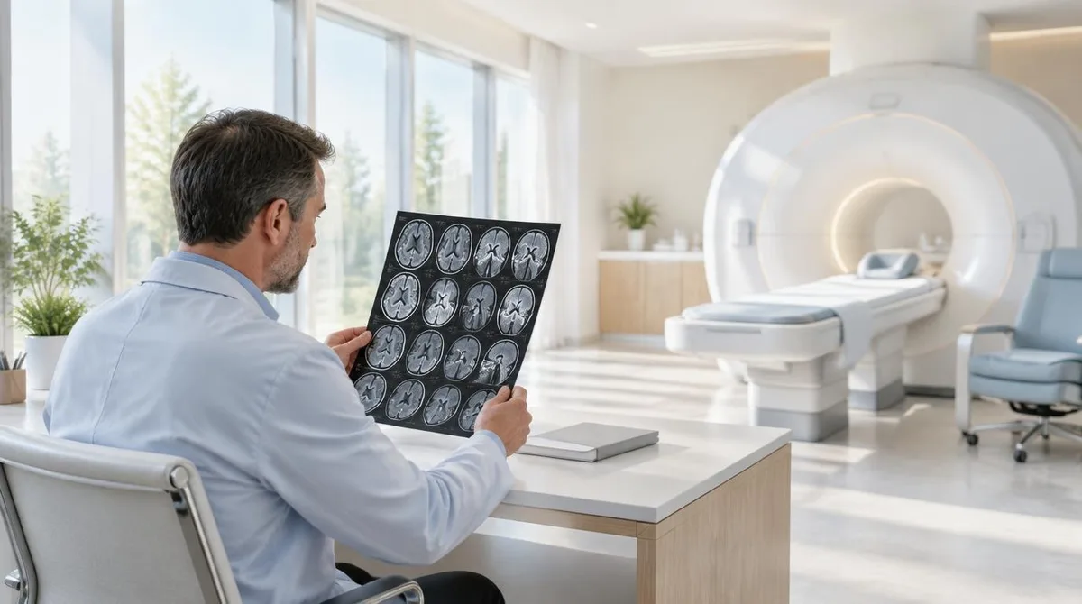
An mri brain scan produces detailed pictures of the brain's soft tissue without using any ionizing radiation. According to the National Institutes of Health, it can tell white matter from gray matter and can help identify aneurysms and tumors. A typical exam runs 30 to 60 minutes, and its value depends on the question being asked: structure, activity, or chemistry.
The method matters because the brain is mostly water and soft tissue, which is exactly what MRI images best. The NIH notes that the brain, spinal cord, nerves, muscles, ligaments and tendons appear much more clearly with MRI than with X-rays or CT. That contrast advantage, plus the absence of radiation, is why MRI is often the modality of choice when frequent imaging is needed, especially in the brain.
This guide explains the physics in plain terms, walks through the main types of brain MRI, and describes what a scan can and cannot show. For readers comparing modalities, the site's imaging technology coverage includes a closer look at how MRI and CT machines each build their image. This connects to our earlier piece, MRI or CT Scan: The Difference Comes Down to Physics and the Question Being Asked.
How does an MRI scanner build an image of the brain?
The scanner aligns hydrogen protons in body water with a strong magnetic field, then reads the energy they release. As the NIH describes it, a low-energy radiofrequency signal tips the protons out of equilibrium; when the field is switched off, sensors detect the energy released as the protons realign. The timing and amount of that energy differ by tissue, which is what allows the computer to distinguish gray matter from fluid, tumor from healthy tissue.
Per the Cleveland Clinic, the machine pairs a temporary magnetic field created by current in coiled wires with radio waves from a transmitter and receiver. The signals the atoms produce are processed into detailed digital images. No X-rays are involved at any step, which is the core safety distinction from CT.
The physics also explains the experience. Protons must be nudged repeatedly, in timed sequences, so the scanner makes loud knocking and clicking sounds for several minutes at a stretch. Patients get earplugs or headphones, and staying still is essential because motion blurs the image.
What does a structural brain MRI show?
A structural scan shows anatomy: the brain's folds, fluid-filled spaces, and the contrast between tissue types. The NIH lists aneurysms and tumors among the conditions MRI can help diagnose in the brain, alongside its ability to separate white matter from gray matter. Radiologists also use it to spot inflammation, bleeding and other changes in soft tissue. For related coverage, see MRI vs CT Scan: How Each Machine Builds Its Image of the Body.
Contrast dye can sharpen the picture. Injected through an IV, contrast helps highlight tumors, inflammation, blood vessels and blood flow, per the Cleveland Clinic, which calls serious reactions rare. The NIH adds one kidney-related caution: the contrast agent gadolinium can raise the risk of a rare disease in people with advanced kidney disease, so patients may first need a blood test to check kidney function.
What a structural scan does not do is read minds or predict disease in a healthy person. It documents anatomy at a moment in time. Subtle findings can be ambiguous, and an image is one input to a diagnosis a clinician makes, never a verdict on its own.
What is functional MRI, and what does it measure?
Functional MRI, or fMRI, measures brain activity indirectly through blood flow. The NIH explains that fMRI uses the blood's magnetic properties to create real-time images of blood oxygenation changes in specific brain areas, which serve as a proxy for neural activity. Areas working harder tend to receive more oxygenated blood.
The clinical use is mapping before surgery. By identifying regions involved in language, motor function or sensation, doctors can locate critical brain areas ahead of an operation. Researchers use fMRI to study healthy brain function, head injuries, epilepsy and neurodegenerative disorders such as Alzheimer's disease, according to the NIH.
The limitation is the word "proxy." fMRI shows where blood oxygenation changed, not the firing of individual neurons, and its findings are group-level patterns in most research settings. It is a powerful research and planning tool, not a lie detector or a window into thoughts.
What is magnetic resonance spectroscopy?
Magnetic resonance spectroscopy, or MRS, measures chemistry rather than anatomy. The NIH describes it as a noninvasive analytic technique that provides a measure of the concentration of different chemical components within tissue. The same magnet and radio waves that build pictures can instead be tuned to report on metabolites.
That chemical readout complements standard MRI. Per the NIH, MRS is particularly useful in identifying diseases affecting the brain, including tumors, strokes and seizures, because it characterizes tissue rather than merely outlining it. A mass that looks ambiguous on a structural image may have a chemical signature that narrows the differential.
MRS is less familiar to patients because it usually happens inside a regular MRI session, as an extra sequence. The practical takeaway is that "an MRI" is not one product but a family of measurements the same machine can make.
What happens during a brain MRI, and what are the risks?
The exam itself is routine. Patients change into a gown, lie face up on a table that slides into the scanner, and remain still for roughly 30 to 60 minutes while the machine knocks and clicks. The Cleveland Clinic notes the technologist can see and speak with the patient through an intercom, and a call button stays in hand.
Preparation is mostly about metal. Because strong magnets interfere with metal objects and implanted devices, the Cleveland Clinic advises telling providers about pacemakers or defibrillators, cochlear implants, aneurysm clips and coils, implanted drug pumps, metallic joint prostheses, stents, and any metal fragments in the body. Jewelry, watches, credit cards, hearing aids and similar items stay out of the exam room because they can interfere with the machine or cause burns.
The main risks are practical rather than radiological. The Cleveland Clinic lists claustrophobia and anxiety, discomfort from lying still, and the noise; open MRI machines with more spacious designs exist for patients who struggle in enclosed spaces. Mild contrast side effects, such as nausea, feeling warm or a metallic taste, occur on very rare occasions. Patients who are pregnant or might be pregnant should tell their provider before contrast is used.
Our analysis: where the method is heading
The throughline across the NIH's overview is that MRI keeps expanding what one magnet can measure: anatomy, blood flow, and now tissue chemistry through MRS, all without radiation. That is why the modality dominates both clinical neurology and cognitive neuroscience research, even though MRI equipment costs more than X-ray or CT, a trade-off the NIH states plainly.
For the research reader, the open question is resolution of meaning. fMRI can map which regions engage during a task, and MRS can report metabolite concentrations, but neither yet delivers a direct readout of the neural events themselves. The gap between what the scanner measures and what the brain is doing remains the field's central measurement problem, and it is where new tracers, higher-field magnets and analysis methods compete. Readers tracking that work can follow the site's research watch coverage and its broader neuroscience section.
What the evidence establishes is durable: MRI images soft tissue without ionizing radiation, differentiates brain tissue types, supports diagnosis and treatment monitoring, and offers functional and spectroscopic variants built on the same physics. What it does not establish is any single-scan conclusion about a person's future. Interpretation stays with qualified clinicians, and the modality's job is to give them better measurements to interpret.