MRI and CT scans both produce detailed pictures of the inside of the body, but they get there in completely different ways. A CT scan uses X-rays taken from many angles. An MRI uses a strong magnetic field and radio waves, with no ionizing radiation at all. That single physical difference drives most of the others: scan speed, image detail, cost, safety profile, and which clinical questions each machine can answer.
The choice between them is rarely about which scan is "better." As MD Anderson neuroradiologist Melissa Chen, M.D., puts it, comparing them is "like comparing apples to oranges" — they are complementary tools that give different kinds of information. A doctor picks the scan that answers the specific question at hand.
This explainer covers how each machine actually works, what it measures well, what the procedure feels like, and where the risks sit. For a decision-focused comparison, the site's earlier piece on CT Scan vs MRI: What Each Machine Measures and When Doctors Choose It covers the ordering logic in more depth. For related coverage, see CT Scan vs MRI: What Each Machine Measures and When Doctors Choose It.
How does a CT scan build an image?
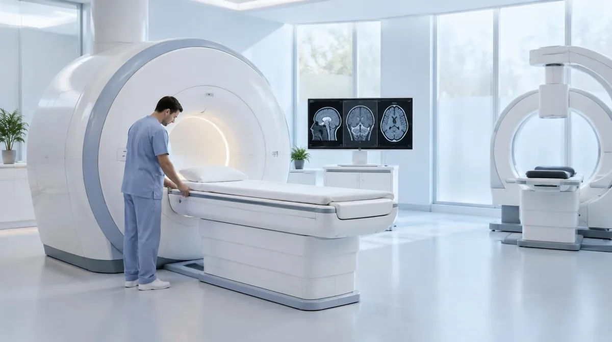
CT stands for computed tomography. The machine fires X-ray beams through the body from multiple angles while the patient lies on a table that slides through a large ring. A computer combines those cross-sectional images into a three-dimensional picture. MD Anderson describes it as a series of X-rays taken very quickly in a circle around the patient.
Because X-rays are absorbed differently by bone, soft tissue, and air, CT is strong at showing edges and structure. Radiologists call this spatial resolution — the ability to show where one structure ends and another begins. That is why CT excels at bone detail, lung imaging, detecting internal bleeding, and emergency work, where speed matters more than fine tissue contrast. Healthline notes a CT typically takes five to ten minutes, and MD Anderson says the scan itself can last a minute or less, with preparation often taking longer than the imaging.
How does an MRI build an image?
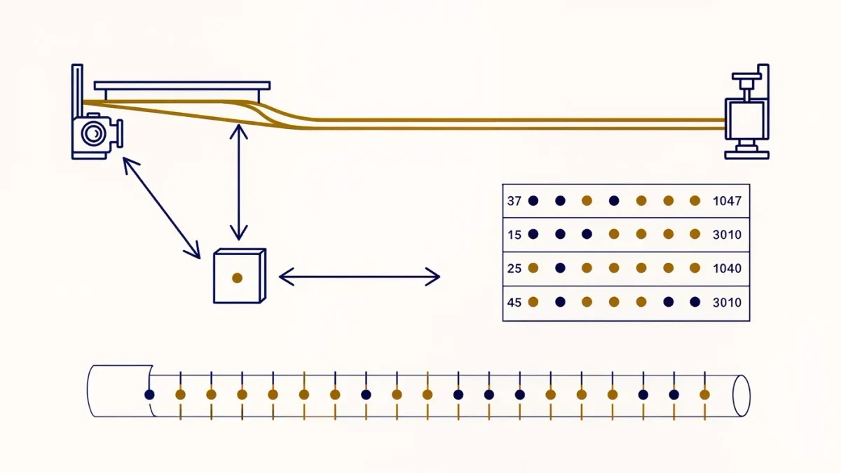
MRI — magnetic resonance imaging — works on an entirely different principle. The machine creates a constant, powerful magnetic field that aligns hydrogen atoms in the body's water and fat molecules. Radio pulses then knock those atoms out of alignment, and the signals they emit as they return to position are translated into an image. No X-rays are involved.
The payoff is contrast resolution: the ability to distinguish between tissue types, fluid, fat, and abnormal growths. mri.md describes MRI as excelling at differentiating soft tissues with extraordinary detail. That is why it is the preferred tool for brain and spinal cord imaging, torn ligaments, herniated discs, and conditions such as multiple sclerosis, where the question is not where a structure is but what kind of tissue it is made of.
The trade-offs are practical. An MRI takes far longer — typically 20 to 90 minutes, according to Healthline — and the patient must lie very still, because movement distorts the images. The scanner is also very loud, so patients are usually offered earplugs or headphones.
What are the safety differences?
The risk profiles differ because the physics differs.
- CT uses ionizing radiation. The dose varies by scan type, and mri.md estimates a single scan's radiation as roughly equivalent to one to ten years of natural background exposure, depending on the scan. It also flags that children are more radiation-sensitive, so MRI is preferred when appropriate.
- MRI uses no radiation, but its magnet creates different hazards. Patients must declare any metal implants — pacemakers, some joint replacements, cochlear implants, or embedded foreign bodies such as shrapnel — because ferromagnetic material can move inside the body. MD Anderson specifically warns about metal fragments from metalworking or injury.
Both scans can involve contrast agents. MRI uses gadolinium-based dyes; CT uses iodine-based ones. Healthline notes that contrast may not be safe for people with kidney disease, and that iodine-based contrast is unsuitable for people with an iodine allergy.
What does each scan cost and how available is it?
CT scans are more common, faster, and generally cheaper. mri.md puts typical costs at roughly $300 to $1,500 for a CT versus $500 to $3,000 or more for an MRI, and notes CT is widely available in emergency rooms around the clock, while MRI access is less universal, especially off-hours. Insurance coverage can also influence which test is approved.
Availability shapes clinical practice in a second way: in an emergency, the faster machine is often the first machine. A common pattern, described by Healthline, is a CT scan first when the cause of symptoms is unclear, followed by an MRI if doctors need a closer look at a specific area.
Which scan is used for which problem?
The split follows directly from what each machine measures best.
- CT is preferred for emergencies such as trauma and suspected internal bleeding, bone fractures, lung imaging, kidney stones, and cancer staging of the chest, abdomen, and pelvis, according to mri.md. It also often guides needle biopsies.
- MRI is preferred for brain and spinal cord questions — tumors, multiple sclerosis, herniated discs — and for joints and soft tissue such as ligament and tendon tears, per the same source. MD Anderson adds that MRI is the preferred scan for brain tumors and the spinal canal, and calls it a "problem-solving tool" for ambiguous CT findings.
Pregnancy changes the calculus too. Medical News Today reports that CT scans and X-rays may not be safe during pregnancy, so doctors may recommend MRI or ultrasound instead — though they may still avoid MRI as a precaution, especially in the first trimester.
Our analysis: the question determines the machine
Read side by side, the sources converge on one point. Neither scan is the superior instrument in any general sense; each answers a different class of question. CT answers where quickly — where the bleed is, where the fracture runs, where the stone sits. MRI answers what carefully — what kind of tissue this is, whether it looks abnormal, how far a lesion extends. The cost, speed, and radiation differences are downstream of that physics, not arbitrary.
For patients, the practical takeaways are limited and well supported: disclose all implants before an MRI, mention a history of many CT scans to a doctor who may weigh MRI alternatives, and expect the MRI room to be loud, long, and cold — MD Anderson explains the cooling systems require it, and heated blankets are offered. The remaining open question for the field is not MRI versus CT at all, but how each machine's measurements map onto specific diseases — which is where molecular tracers and advanced sequences, the usual subjects of this publication, come in.